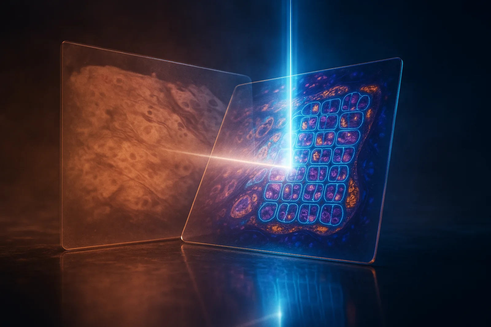
Slides do not politely stay aligned.
A pathology lab may scan an H&E slide for tissue architecture, an IHC slide for protein expression, a PAS slide for renal structure, and a multiplex immunofluorescence slide for cellular markers. The human story is that these images come from the same biopsy. The computational story is less sentimental: the tissue has been sliced, stained, bleached, re-stained, stretched, torn, folded, scanned, and generally treated like a fragile biological object in a world built for rectangles.
So when a downstream AI system wants to compare marker expression with tissue morphology, the first problem is not diagnosis. It is geography.
CORE, short for Cell-level Coarse-to-fine Registration Engine, is a new registration framework for aligning multi-stain whole-slide images across tissue and nuclei scales.1 Its contribution is not that it “does pathology”. It does not classify tumours, predict survival, or whisper diagnostic wisdom into a consultant’s ear. It performs the less glamorous task that makes many of those ambitions technically possible: it tries to put different stained versions of tissue into a shared coordinate system.
That distinction matters. In digital pathology, image registration is infrastructure. And like most infrastructure, nobody notices it until it fails.
CORE treats alignment as a two-speed problem
The useful idea in CORE is not one clever module. It is the division of labour.
The paper argues that whole-slide image registration needs two different kinds of intelligence. First, the system must align the tissue globally, despite stain differences and large physical deformations. Then, if the application requires cellular precision, it must refine the alignment using nuclei as biological landmarks.
That gives CORE its coarse-to-fine structure:
| Stage | Scale | Main mechanism | Operational purpose | Cost profile |
|---|---|---|---|---|
| Coarse registration | Low magnification | Prompt-based tissue masking, TriMorph, XFeat feature matching, non-rigid deformation | Fast global slide alignment | Seconds per pair in reported coarse experiments |
| Fine registration | High magnification | Nuclei detection, shape-aware point-set alignment, CPD deformation | Nuclei-level or near-cellular alignment | Tens of minutes per pair in reported fine experiments |
That table is the paper’s practical thesis. Coarse registration is for throughput. Fine registration is for precision. Confusing the two is how procurement decks become tragic little works of fiction.
The coarse stage is designed to solve the “same tissue, different visual language” problem. Different stains highlight different biological structures. H&E, IHC, PAS, and mIF do not merely change colour; they alter which features are visible, prominent, or useful for matching. A method that relies too heavily on intensity similarity can be fooled because the same physical region may look completely different under another stain.
CORE begins by making the tissue easier to isolate. It uses a prompt-based tissue mask extraction step built around Florence-2 and SAM, applying prompts such as tissue, stain, histology, and cell to separate tissue from background and artefacts. When this fails, which the authors report for roughly 2–3% of WSIs due to poor quality or staining, the system falls back to a U-Net trained on ACROBAT tissue samples.
This is not decorative preprocessing. It narrows the problem from “align two enormous, noisy images” to “align the tissue regions that matter”. At WSI scale, that reduction is not a convenience; it is survival.
TriMorph gets the tissue roughly right before XFeat gets clever
After mask extraction, CORE estimates a rigid transformation using what the paper calls TriMorph. The name is mildly theatrical, but the mechanism is sensible: translation, scale, and rotation are estimated from tissue morphology.
The centre of mass of the source tissue mask is aligned with the target. Relative tissue dimensions guide scale adjustment. Candidate rotations are evaluated by tissue overlap, using the Dice coefficient to choose the best alignment. If no transformation passes the threshold, the method returns an identity transform rather than hallucinating a confident mistake. A small mercy, but a useful one.
TriMorph gives the system a physically plausible starting point. XFeat then performs accelerated dense feature extraction, detecting and matching up to 16,000 keypoints between downsampled WSI pairs. The key point here is not that XFeat is magical. It is that CORE uses deep features where they are operationally appropriate: to capture stain-invariant structural cues at a coarse level, not to pretend that colour-normalised pixels are enough.
The coarse pipeline then estimates non-rigid deformation through a multiresolution pyramid. It uses local normalised cross-correlation as the similarity metric and smoothness regularisation to avoid implausible displacement fields. The result is a deformation model that can handle broad tissue warping without immediately diving into the full-resolution cellular swamp.
The authors report that the best coarse-level performance appears at 0.625× and 1.25× objective resolutions. That is a useful engineering observation. It says the right low-resolution view can preserve enough tissue structure for alignment while avoiding the computational punishment of full-resolution processing.
The nuclei stage is where CORE becomes biologically specific
The fine stage starts after coarse alignment. CORE detects nuclei in both the target slide and the coarsely registered source slide, then treats them as point sets.
Here the paper makes a deliberately unfashionable choice: it uses a morphology-based nuclei detection pipeline rather than a deep learning nuclei detector. The reason is practical. Deep learning nuclei models often require annotated nuclei data tuned to tissue type, stain, scanner, and segmentation objective. Most registration workflows do not have that luxury. CORE instead uses greyscale conversion, adaptive thresholding, H-maxima and H-minima operations, marker-controlled watershed segmentation, and centroid extraction.
In other words, it chooses portability over maximal segmentation elegance. This is exactly the sort of decision that rarely excites conference audiences but often determines whether a method survives contact with hospital data.
The shape-aware alignment step then refines global transformation using both spatial proximity and morphological similarity. Each nucleus is not merely a coordinate; it also carries a shape attribute such as area. The hybrid distance metric weights morphology with a parameter set to 0.3 in the authors’ evaluation. Too little shape information and the method behaves like ordinary point matching. Too much and it risks overfitting to morphology. The paper positions 0.3 as a compromise between spatial alignment and biological plausibility.
For re-stained slides, CORE can use the complete detected nuclei set because the underlying tissue section is physically the same. For consecutive sections, the problem is harder: neighbouring slices are similar, not identical. The authors therefore use progressive sample size scaling, beginning with 500 nuclei and doubling up to 200,000, selecting the transformation with the lowest final error. They report that accuracy stabilises at intermediate sample sizes around 150,000 points.
That observation is important because it undercuts a lazy assumption: more nuclei are not automatically better. At some point, the algorithm is paying extra rent for almost no additional accuracy.
Finally, CORE applies Coherent Point Drift for non-rigid point-set registration, using mutual nearest neighbours to enforce bidirectional consistency. The resulting displacement field is interpolated, smoothed, constrained by maximum displacement, and checked using the Jacobian determinant to avoid local folding. Regions with non-positive Jacobian determinant are zeroed out.
That final detail matters. A deformation field can look numerically satisfying while being anatomically absurd. CORE at least checks whether the transformation has started folding tissue through itself, which is generally considered impolite.
The evidence is broad, but not all evidence does the same job
CORE is evaluated on three public datasets and two private cohorts: ACROBAT, ANHIR, HYRECO, Multi-IHC CRC, and REACTIVAS. The evidence is not a single leaderboard claim. It is a bundle of tests serving different purposes.
| Evidence source | Likely purpose | What it supports | What it does not prove |
|---|---|---|---|
| ACROBAT | Main coarse-registration comparison | CORE is highly competitive and faster on large breast cancer WSI alignment | Fine nuclei-level performance, because ACROBAT was available at 10× and fine registration was not run |
| ANHIR | Public benchmark across tissue and stain diversity | CORE improves average rTRE, with fine registration improving accuracy further | That fine registration is always worth the runtime for every workflow |
| HYRECO | Consecutive and re-stained section comparison | CORE performs well where physical section similarity varies | Universal performance across all tissue types or cutting protocols |
| Multi-IHC CRC | Private validation on colorectal multi-IHC slides | CORE transfers beyond public benchmarks and improves coarse and fine rTRE | Broad clinical generalisation, since the evaluation subset is limited |
| REACTIVAS | Private multimodal renal biopsy extension with H&E, PAS, and mIF | CORE can handle bright-field and immunofluorescence combinations | Full deployment readiness for renal pathology workflows |
| Appendix timing and stepwise tables | Implementation and diagnostic evidence | Where the runtime goes and how each stage reduces error | A complete ablation of every design choice |
That last row deserves emphasis. The appendix is useful, but it is not a second thesis hiding in the furniture. It gives parameter settings, stepwise error reductions, and processing time by module. It helps interpret the method. It does not prove that every hyperparameter is globally optimal.
The numbers say “faster coarse, better fine”, not “free precision”
The headline results are strong, but the pattern is more interesting than the victory lap.
On ACROBAT, CORE’s coarse method reports an AMTRE at the 90th percentile of 139.00 μm with a mean time of 14.37 seconds. DeeperHistReg reports 140.33 μm at 70 seconds, HistokatFusion 141.64 μm at roughly 50 seconds, and NEMESIS 209.76 μm at 120 seconds. This is the cleanest coarse-stage business case: similar or better accuracy with materially lower runtime.
ANHIR shows the price of cellular ambition. CORE’s coarse method reports an average rTRE of 0.0040 at 12 seconds. Fine CORE improves the average rTRE to 0.0034, but runtime rises to 1,680 seconds. The median metrics also improve, but one maximum-error metric worsens from 0.0240 to 0.0323. That does not invalidate the fine method. It simply means fine registration is not a magic universal reducer of every tail error. Imagine that: biology remains annoying.
HYRECO is where the re-stained versus consecutive distinction becomes especially useful. For consecutive sections, CORE reports a final median TRE of 4.35 μm, compared with 4.96 μm for DeeperHistReg and 5.30 μm for HistokatFusion. For re-stained sections, CORE reports 0.41 μm, compared with 0.59 μm for DeeperHistReg and 0.90 μm for HistokatFusion. The result aligns with intuition: re-stained slides preserve the same tissue section, so nuclei-level correspondence is more meaningful than in neighbouring slices.
On Multi-IHC CRC, CORE’s coarse method reports AArTRE of 0.00431 at 14.37 seconds, while fine CORE improves this to 0.002281 at 2,172 seconds. On REACTIVAS, the coarse method reports AMTRE of 1.0272 μm for PAS and 2.0495 μm for mIF, improving to 0.36 μm and 0.8460 μm respectively after fine registration, with fine runtime reported at 1,500 seconds.
The operational interpretation is plain: CORE buys speed at the tissue level and precision at the nuclei level, but it does not abolish the trade-off. Fine registration is valuable when the downstream task actually needs cell-level correspondence. If a workflow only needs broad tissue alignment, running the full fine stage may be computational theatre.
The business value is workflow leverage, not diagnostic magic
The likely misconception is that CORE is another diagnostic AI model. It is not. It is a registration engine. That makes it less flashy and more strategically interesting.
In a multi-stain pathology workflow, registration determines whether signals from different slides can be compared reliably. Poor alignment can corrupt biomarker correlation, spatial phenotyping, microenvironment analysis, and multimodal model training. If the tissue map is wrong, the downstream model may still produce confident outputs. It will just be confidently analysing a coordinate system held together with optimism and masking tape.
CORE’s business relevance depends on the workflow:
| Workflow need | CORE stage that matters most | Business interpretation |
|---|---|---|
| Slide-level comparison across stains | Coarse registration | Faster alignment can reduce manual checking and preprocessing bottlenecks |
| Biomarker correlation across IHC or PAS slides | Coarse plus selective fine registration | Use fine alignment where regions of interest require higher precision |
| mIF and bright-field integration | Coarse-to-fine pipeline | Enables richer multimodal tissue maps, especially for research and translational studies |
| Spatial phenotyping or single-cell analysis | Fine nuclei-level registration | Precision may justify runtime when cell correspondence changes the scientific answer |
| Routine high-throughput production | Mostly coarse registration, with triage | Fine registration must be rationed unless runtime is reduced |
For hospitals, the near-term benefit is not replacing pathologists. It is making multimodal slide workflows less manually brittle. For biopharma and translational research groups, the benefit is stronger: multi-stain and mIF studies often live or die by the ability to compare spatial signals across sections. If CORE can reduce alignment friction while preserving biological correspondence, it becomes part of the data plumbing for biomarker discovery and tissue microenvironment analysis.
For AI developers, CORE is a reminder that better models often require better coordinates before they require bigger networks. The paper also provides open-source code and TIAViz visualisation support, which matters because registration results need visual inspection. A lower TRE is nice; a deformation field that a human can inspect is better.
The runtime table reveals the real bottleneck
The appendix timing table is one of the more practically useful parts of the paper. Coarse operations are cheap. Tissue mask extraction takes around 5.40 seconds on GPU. TriMorph takes 0.56 seconds. XFeat takes 0.80 seconds. Warping is effectively negligible.
The fine stage is different. Fine point-set rigid registration takes 299 seconds on GPU, and fine point-set non-rigid registration takes 400 seconds. The paper’s dataset-level fine runtimes are higher still, ranging around 1,500 to 2,850 seconds depending on the experiment.
This is where business users should resist the temptation to average everything into one vague “registration time”. CORE has two products inside it. One is fast coarse alignment. The other is slower precision alignment. Buying the second for every slide pair may be justified in a spatial biology lab. It may be wasteful in a routine workflow where tissue-level overlap is enough.
A sensible deployment would probably use tiering:
- Run coarse registration by default.
- Score whether the downstream task needs nuclei-level precision.
- Apply fine registration only to selected slides, regions, or research workflows.
- Use visual inspection tools to audit high-value cases.
- Track failure modes by stain pair, tissue type, scanner, and sample preparation protocol.
That is less glamorous than “end-to-end AI”. It is also how serious systems are built.
The boundaries are specific, not ceremonial
CORE’s limitations are not generic “more research is needed” wallpaper. They affect deployment choices.
First, fine registration depends on nuclei detection. Overlapping cells, staining artefacts, poor segmentation, and dense tissue regions can undermine the point sets that drive alignment. The authors choose classical nuclei detection partly to avoid dataset-specific annotation requirements, but that choice also means performance will vary with image quality and tissue morphology.
Second, consecutive sections are not re-stained sections. Re-stained slides preserve the same physical tissue section; consecutive slides do not. A nuclei-level method can improve local alignment for consecutive sections, but it cannot create biological structures that are absent from one slice. Some “misalignment” is actually biology and sectioning, not algorithmic failure.
Third, some evaluations are necessarily narrower than the headline dataset list suggests. ACROBAT is large, but fine registration was not run because the slides are available at 10×. Multi-IHC CRC uses a selected evaluation subset of seven cases with six slides per case. REACTIVAS includes 11 paired renal biopsy samples across H&E, PAS, and mIF. These are valuable tests, especially because they cover difficult modalities, but they are not proof of universal deployment performance.
Fourth, the paper evaluates registration accuracy and visual alignment, not downstream clinical outcomes. CORE may enable better diagnostic or research pipelines, but the paper does not show that it improves diagnosis, treatment selection, trial enrolment, or patient outcomes. Those are downstream claims requiring separate validation.
Finally, the method’s future acceleration is not free. The authors note that faster deep learning models for nuclei detection could reduce runtime, but such models may require explicit training or fine-tuning for each dataset. That is the familiar trade: automation improves, and then someone has to feed it annotations. The machine is efficient; the humans are merely invoiced elsewhere.
CORE’s real lesson is architectural discipline
The strongest part of CORE is its refusal to solve every subproblem at the same scale.
At low resolution, it uses tissue masks, morphology, deep feature matching, and smooth deformation to get the whole slide into approximate agreement quickly. At high resolution, it changes representation: nuclei become points, shape becomes a cue, and deformation becomes a local biological mapping problem.
That is the right instinct for computational pathology. Whole-slide images are not just big photographs. They are multiscale biological records. A method that treats global tissue alignment and nuclei-level correspondence as the same problem is usually either too slow, too brittle, or too impressed with itself.
CORE is not the final word on WSI registration. Its fine stage is expensive. Its nuclei detection can fail where tissue quality is poor. Its private cohorts are limited. Its clinical value remains indirect until downstream applications are validated.
But as infrastructure, it is persuasive. It shows that multistain registration can be made both faster and more biologically grounded when the pipeline respects scale. For organisations building digital pathology, spatial biology, or multimodal tissue analytics systems, that is the useful lesson: before asking AI to interpret the tissue, make sure the tissue is in the same place.
Astonishingly, coordinates still matter.
Cognaptus: Automate the Present, Incubate the Future.
-
Esha Sadia Nasir et al., “CORE - A Cell-Level Coarse-to-Fine Image Registration Engine for Multi-stain Image Alignment,” arXiv:2511.03826, 2025. https://arxiv.org/abs/2511.03826 ↩︎