Pretty in Pink Is Not Enough: Virtual 3D H&E Needs Structural Proof
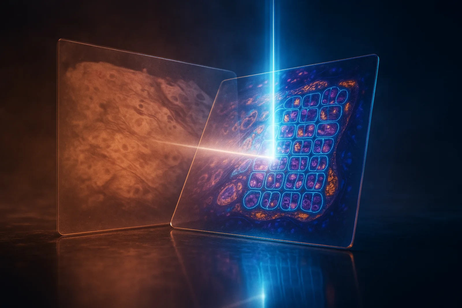
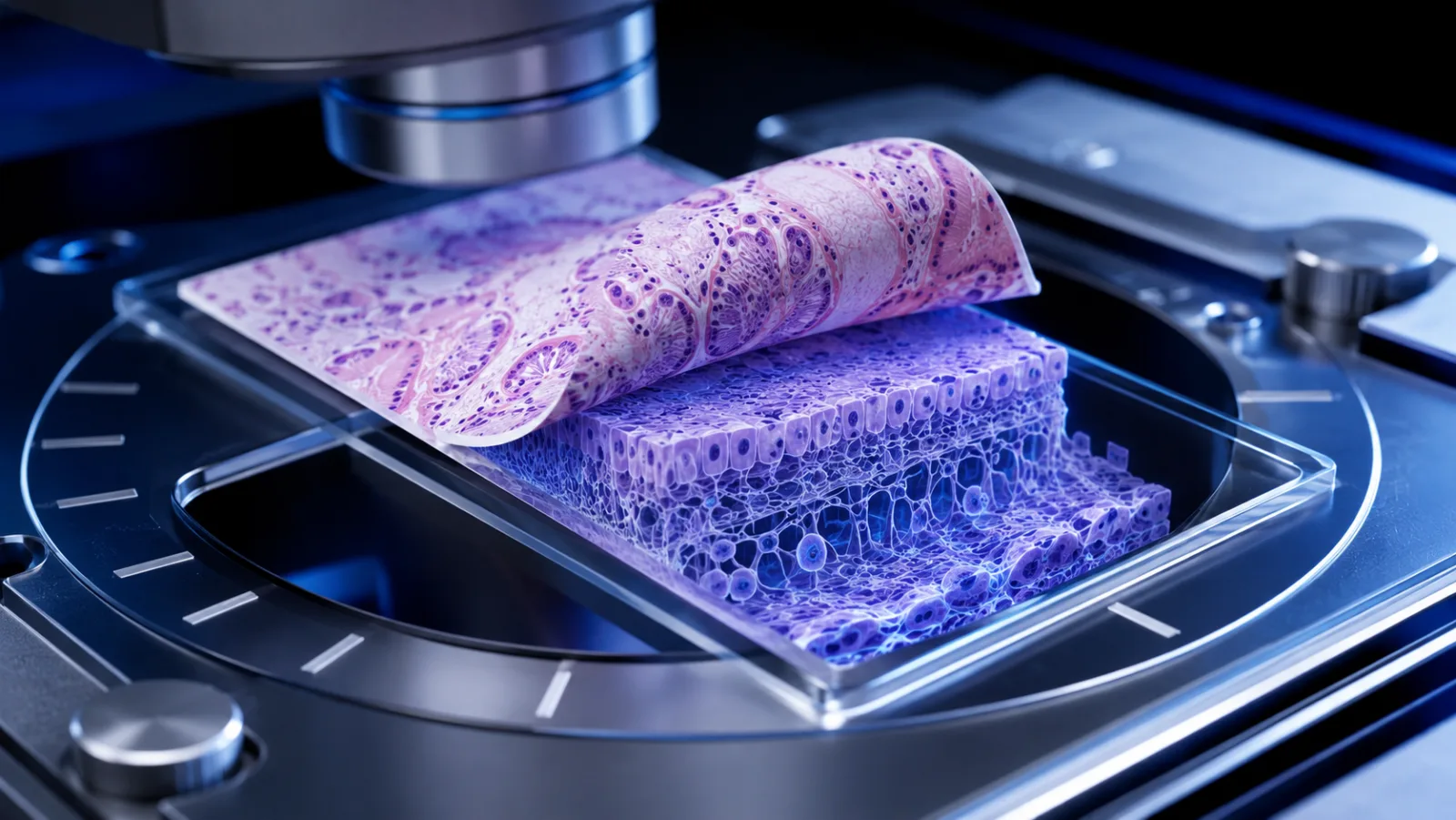
TL;DR for operators The useful part of this paper is not that it makes label-free microscopy look like H&E. That is the easy headline, and also the easiest way to misunderstand the work. The paper introduces HistoBIT3D, a dataset that pairs phase-contrast Back-illumination Interference Tomography, or BIT, with voxel-wise registered fluorescence-labelled nuclei in 3D tissue volumes.1 That matters because virtual staining has a basic governance problem: a generated image can look histological while quietly moving, deleting, or inventing cellular structure. In pathology, that is not a charming hallucination. It is the sort of thing that gets written up after the incident review. ...