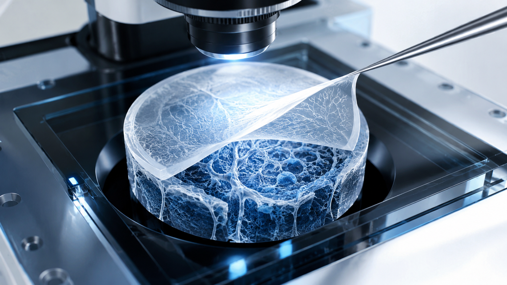
Microscopy has a labor problem.
Not the photogenic kind where a scientist leans into a glowing instrument and discovers the secret architecture of life before lunch. The duller problem is that modern light sheet fluorescence microscopy can produce rich three-dimensional volumes faster than expert teams can label them. Segmentation requires voxel-level masks. Stain classification requires domain knowledge. Restoration needs paired degraded and high-quality images, which nature, unhelpfully, does not always provide in tidy folders.
So the real bottleneck is not whether an AI model can be trained. It is whether a model can become useful before the annotation bill eats the project.
That is the business-relevant question behind arXiv:2605.26026, where Scheinfeld and colleagues introduce a multimodal 3D foundation model for light sheet fluorescence microscopy, or LSM.1 The paper is not best read as “another model beats baselines on three tasks.” That summary is technically true and editorially sleepy. The more important mechanism is this: the authors try to convert unlabeled volumetric microscopy data, plus lightweight biological language descriptions, into a reusable representation that can later be adapted with much smaller labeled datasets.
That mechanism matters because LSM is exactly the sort of domain where standard supervised AI logic becomes expensive. The images are large, three-dimensional, biologically diverse, and often tied to staining protocols that general-purpose vision models do not understand. A model trained for one structure may not transfer cleanly to another. A 2D method may slice through the data and miss the axial continuity that makes 3D imaging valuable in the first place. A universal “segment anything” promise sounds wonderful until the object is not a standard object, the background is biological tissue, and the thing being segmented is a faint vessel network or sparse amyloid plaque.
The paper’s contribution is therefore not that it magically solves microscopy. It is that it gives a concrete answer to a narrower and more operational question: can a 3D LSM-specific pretraining pipeline make downstream microscopy analysis less dependent on large annotated datasets?
The model is built around missing evidence, not around labels
The pretraining setup starts from a useful premise: LSM labs often have more raw volumes than labels. The authors assemble 1,023 volumetric 96³ voxel patches from internal and public sources, spanning multiple organisms, structures, stains, and imaging protocols. The internal component includes 24 single-channel mouse whole-brain images, each with a distinct immunostain. Public data come from SELMA3D and Allen Institute resources. This is not internet-scale pretraining, but in 3D microscopy, even a thousand curated volumetric patches are not a trivial object. Each patch carries spatial structure, staining behavior, and imaging artifacts that a 2D model may flatten away.
The model then learns from a deliberately damaged version of the image. A student network receives a masked 3D input volume, where random non-overlapping patches are zeroed out. A teacher network sees the unmasked volume. The teacher is not updated by ordinary gradient descent; its weights are maintained as an exponential moving average of the student. In plain English, the student is asked to infer stable volumetric representations from partial evidence, while the teacher provides a smoother full-context target.
This is the first mechanism worth keeping in mind. The model is not merely memorizing labels. It is learning what biological volume structure tends to look like when some evidence is hidden. For segmentation and deblurring, that is a relevant pretraining discipline. A vessel does not stop being a vessel because one slice is noisy. A cell nucleus does not become a new class because a small patch is masked. The model is being trained to treat missing local evidence as a reconstruction problem inside a larger 3D context.
The paper combines four loss components:
The first two terms are image-only: distillation from the teacher’s feature distribution and voxel-level reconstruction of masked regions. The second pair brings in language: cosine alignment between image and text embeddings, and a CLIP-style contrastive loss that pulls matching image-text pairs together while pushing mismatched pairs apart.
That split is important. The framework can run without text by disabling the alignment and contrastive terms. The paper therefore gives us a useful contrast: image-only pretraining versus image-plus-text pretraining. It is not a perfect loss ablation, but it is the paper’s most practical test of whether biological language adds value beyond volumetric structure.
The text branch gives semantic hints, not supernatural understanding
The captions in this paper are not random alt text scraped from the web. Domain experts compose structured two-to-four sentence descriptions for each volume. These captions include staining target, imaging channel, organism and tissue source, visible morphology, spatial organization, and relevant pathology. The authors then use an LLM to paraphrase captions for linguistic diversity while trying to preserve meaning.
This is a careful design choice, and also a useful boundary. The text branch is not replacing microscopy expertise. It depends on it. The model receives biological language because experts have already translated visual and experimental context into words. The LLM paraphrasing step may increase linguistic variety, but the scientific signal originates in expert captioning.
For business readers, the implication is straightforward: multimodal pretraining does not remove the need for domain knowledge. It changes where that knowledge is used. Instead of paying experts only to label every downstream task from scratch, a platform may ask experts to help build reusable pretraining metadata and then spend fewer labels on each later task. That is a potentially better capital allocation. It is not a free lunch. More like a lunch where the invoice arrives earlier, under a less annoying cost center.
The image-only results also keep the story honest. Across the paper, structural cues alone are already powerful. In several settings, the image-only model is competitive with, or better than, the image-plus-text variant. That does not make language useless. It means text is complementary, not magical. The strongest interpretation is not “multimodal always wins.” It is “3D LSM-specific pretraining helps, and language can help in selected regimes.”
That distinction matters because the easiest bad reading of this paper is to treat it as a universal biology foundation model. It is not. It is an LSM-focused volumetric representation learner tested on segmentation, stain classification, and synthetic deblurring. The ambition is reusable microscopy analysis, not automatic biological interpretation on command.
The downstream tasks are evidence for transfer, not three separate product demos
The paper evaluates three downstream tasks: voxel-wise binary segmentation, patch-level stain classification, and deblurring. A weaker article would summarize them one by one and then declare victory. A better reading asks what each task tests about the pretrained representation.
| Test | Likely purpose in the paper | What it supports | What it does not prove |
|---|---|---|---|
| Segmentation on amyloid plaque, cell nucleus, and vessels | Main evidence for annotation-efficient spatial understanding | The pretrained backbone can improve voxel-level prediction with only 5 or 15 training patches per datatype | It does not prove universal segmentation across all biological structures or acquisition protocols |
| Classification across 12 stain categories | Main evidence for semantic transfer | The representation captures stain- and morphology-relevant features better than PCA, ResNet-18, and scratch models in the tested setup | It does not prove open-ended biological diagnosis or zero-shot stain recognition |
| Deblurring with synthetically blurred inputs | Exploratory downstream extension and restoration evidence | Pretrained features can help reconstruct cleaner 3D volumes under synthetic degradation | It does not establish performance on all real microscope blur, motion artifacts, or acquisition failures |
| Image-only versus image-plus-text variants | Ablation-like comparison | Structural pretraining is strong; language adds value in some cases | It does not isolate every loss term independently |
| Overtrained SwinUNETR variant | Robustness/sensitivity test around pretraining duration | Standard pretraining is broadly sufficient, with modest gains in selected settings from much longer exposure | It does not prove that more pretraining data or epochs will scale monotonically |
| Blinded expert ranking of segmentation outputs | External qualitative validation | Metric gains correspond to perceptible quality differences for experts | It does not replace prospective biological or clinical validation |
The segmentation test is the cleanest fit with the paper’s thesis. The authors use three datatypes from SELMA3D: amyloid plaques, cell nuclei, and vessels. These are not interchangeable shapes. Amyloid plaques are sparse and punctate. Cell nuclei are dense foreground objects. Vessels are elongated, connected structures where continuity and topology matter. That makes segmentation a reasonable stress test for whether the representation learned something more general than one object class.
In the few-shot segmentation setting, the model trains with only five patches per datatype. In the many-shot setting, it trains with 15. Results are averaged across held-out patches from three cross-validation folds. The comparison includes task-specific baselines such as µSAM, CellSeg3D, and Cellpose-SAM variants, plus models trained from scratch.
The numerical pattern is not perfectly uniform, which is precisely why it is useful. For amyloid plaque segmentation, the image-plus-text UNet reaches a few-shot total Dice of 0.68 and instance Dice of 0.58, versus scratch UNet at 0.50 and 0.11. That is not a cosmetic gain; instance Dice moving from near-failure to materially useful is the kind of result that annotation-constrained teams notice. In the many-shot amyloid setting, the same model reaches 0.80 total Dice and 0.69 instance Dice, while the overtrained Swin variant reaches 0.83 instance Dice.
For nuclei, scratch models are already strong, especially on instance Dice. The improvements are therefore more modest and architecture-dependent. For vessels, the strongest pretrained variants reach around 0.92 total Dice in the many-shot setting, ahead of scratch and µSAM baselines in the reported table, but the gap is smaller than the amyloid instance result. That pattern suggests the business value will not be evenly distributed. The biggest payoff appears where low-label learning is genuinely hard for scratch models, not where the supervised baseline is already comfortable.
Classification shows semantic transfer, but the “text wins” story is too simple
The classification experiment expands the task beyond the segmentation/deblurring datasets by adding additional stain categories, producing 12 image categories. The few-shot setting uses 56 training samples, while the many-shot setting uses 105. Baselines include PCA and 3D ResNet-18, along with scratch-trained UNet and Swin variants.
Here the pretrained UNet is the star, but the best pretraining mode depends on the data regime. In the few-shot setting, image-only UNet reaches 0.71 accuracy and 0.69 macro F1, compared with scratch UNet at 0.49 accuracy and 0.46 macro F1, PCA at 0.33/0.28, and ResNet-18 at 0.36/0.27. In the many-shot setting, image-plus-text UNet reaches 0.74 accuracy and 0.69 macro F1, compared with scratch UNet at 0.61/0.57.
This is where the paper becomes more interesting than a marketing abstract. If language were the whole story, image-plus-text would dominate consistently. It does not. In few-shot classification, image-only UNet is stronger than image-plus-text UNet. In many-shot classification, image-plus-text UNet is stronger. Swin variants show their own pattern: image-plus-text Swin is weak in few-shot accuracy but stronger in many-shot classification.
A fair interpretation is that the text branch can enrich representations, especially when the downstream setting can use semantic context without being overwhelmed by optimization noise or small-data instability. But volumetric structure remains the foundation. In LSM, morphology is not a decorative feature; it is the signal.
For business use, this means an AI microscopy platform should not automatically default to “more modalities must be better.” The practical question is task-level: does language metadata improve the downstream metric that matters, under the amount of labeled data actually available? The paper’s answer is “sometimes, materially.” That is more useful than “always,” because “always” tends to become expensive and false.
Deblurring is promising, but synthetic blur keeps the claim narrower
The deblurring experiment asks the model to reconstruct sharp volumes from synthetically blurred inputs. The training objective combines L1 reconstruction, SSIM, a 3D gradient-based edge consistency term, and a high-frequency loss. The reported metric is SSIM, with PSNR said to follow similar trends.
The results are supportive but mixed. For amyloid plaque deblurring, pretrained UNet variants slightly improve over scratch UNet: image-plus-text UNet reaches 0.65 SSIM in few-shot and 0.69 in many-shot, compared with scratch UNet at 0.63 and 0.67. For cell nuclei, image-only UNet reaches 0.86 and 0.89, compared with scratch UNet at 0.81 and 0.87. For vessels, image-only UNet reaches 0.89 and 0.92, compared with scratch UNet at 0.88 and 0.90.
Those are real gains, but they are not the same kind of evidence as the segmentation improvements. Synthetic blur is a controlled degradation. Real microscopy restoration can involve optical blur, scattering, motion, staining variation, acquisition noise, and sample preparation artifacts. Some of those may resemble the synthetic setup. Some will not. The correct reading is that pretrained 3D representations can support restoration tasks; not that the model is ready to repair every failed acquisition.
There is still a business lesson here. Deblurring is not merely a cosmetic add-on. In high-throughput imaging workflows, restoration quality can affect downstream segmentation, quantification, and review time. A representation that transfers to restoration may help build pipelines where acquisition, cleanup, segmentation, and interpretation are less siloed. But the paper’s strongest evidence remains annotation-efficient segmentation and classification, not universal image repair.
The expert evaluation is small, but it answers the right sanity-check question
Metric improvements in biomedical imaging are useful only if they correspond to differences experts can see or trust. Otherwise, one is just polishing decimals in the basement.
The paper includes a blinded expert evaluation of segmentation outputs. Six domain experts, each with five to ten years of LSM experience, independently evaluated 60 segmentation predictions across multiple datatypes. Outputs were ranked best, middle, or worst, scored as 2, 1, or 0. The pretrained image-plus-text UNet achieved an average score of 1.43, compared with 1.07 for scratch UNet and 0.50 for µSAM base.
This is not a large human-factors study, and it does not prove deployment readiness. But it serves an important purpose: it checks whether quantitative gains produce visibly better segmentation quality. The answer, in this tested setting, is yes. That matters because biomedical image analysis often fails in the gap between metric optimization and expert acceptance. A model that improves Dice while producing strange boundaries may still waste expert time. Here, the authors at least test the perceptual side of the claim.
Notice what the expert evaluation does not do. It does not assess downstream biological discovery. It does not test multi-site prospective deployment. It does not validate clinical decision-making. It is a qualitative external validation layer for segmentation quality. That is valuable, provided we do not ask it to be more than it is.
The business value is reusable representation capital
The paper’s most practical idea is that microscopy organizations may need to think less in terms of one-off model training and more in terms of representation capital.
In a one-off pipeline, every new structure or stain requires a fresh annotation push, model training, validation, and correction cycle. That can work for a narrow lab process. It scales poorly across organisms, tissues, protocols, and tasks. In a representation-capital approach, raw historical volumes and expert captions become assets for pretraining. Downstream teams then adapt the backbone with smaller task-specific labels.
For a lab, core facility, biotech imaging platform, or AI-enabled pathology-adjacent workflow, the ROI logic looks like this:
| Technical contribution | Operational consequence | ROI relevance |
|---|---|---|
| 3D LSM-specific pretraining on heterogeneous volumes | The model starts with volumetric priors closer to the target modality | Less need to force general 2D vision models into 3D biological problems |
| Masked reconstruction and EMA distillation | The backbone learns stable structure from partial evidence | Better low-label transfer for segmentation and restoration-like tasks |
| Expert-authored captions with image-text alignment | Biological semantics can be embedded into the representation | Potential reuse of expert knowledge across tasks, not only per-label annotation |
| Few-shot finetuning across segmentation, classification, and deblurring | One pretrained backbone supports several analysis workflows | Lower marginal cost for new downstream tasks, if validation holds |
| Blinded expert evaluation | Quality is checked against human perception, not only metrics | Better chance of adoption in expert-led workflows |
This is not “AI replaces the microscopist.” That slogan should be retired somewhere quiet. The more serious value proposition is workflow leverage: fewer labels, faster adaptation, and more reusable infrastructure for recurring imaging tasks.
The paper also suggests a governance point. If captions, pretraining data, and fine-tuning datasets are part of the model’s asset base, then data stewardship becomes central. Teams need to know which organisms, stains, tissue types, imaging conditions, and annotation conventions are represented. A model pretrained on one distribution may be excellent inside that distribution and brittle outside it. The inventory of pretraining coverage becomes a business document, not just a methods appendix.
The boundaries are where deployment decisions should begin
The paper is promising, but its boundaries are not minor footnotes. They define where a business interpretation can safely stand.
First, the evaluation scale is limited. The pretraining set contains 1,023 volumetric patches, and the annotated downstream segmentation/deblurring dataset contains 84 patches. The authors use cross-validation and held-out testing, but the absolute number of held-out samples is small. This is normal for specialized biomedical imaging. It is also why the result should be treated as evidence of feasibility, not final proof of broad deployment robustness.
Second, the domain is LSM-centered. The model is designed around light sheet fluorescence microscopy volumes. It should not be casually generalized to all microscopy modalities, all histology, all radiology, or “biology images” as a category. The paper’s strength is domain specificity. Diluting that into a universal claim would make it less accurate, not more impressive.
Third, deblurring uses synthetic degradation. This is useful for controlled evaluation, but real acquisition failures may behave differently. Any restoration workflow would need site-specific testing against real artifacts.
Fourth, the language branch depends on caption quality. Expert-written captions are valuable, but they impose their own cost and bias. LLM paraphrasing may diversify wording, but it does not create new biological truth. If captions are incomplete, inconsistent, or too far removed from visible image evidence, the multimodal component can become decorative or even misleading.
Finally, the paper does not eliminate expert review. It may reduce annotation burden and improve model initialization. It does not remove the need for QA, validation, protocol tracking, or domain oversight. In biomedical workflows, that is not pessimism. It is plumbing.
The takeaway: smaller labels, not smaller responsibility
Scheinfeld and colleagues give a credible early answer to a practical question: how can LSM teams use the unlabeled volumetric data they already produce to reduce the cost of downstream analysis?
The answer is mechanism-first. Train a 3D model to understand volumetric structure by reconstructing masked evidence. Stabilize learning through an EMA teacher. Add biological language when expert captions can provide useful semantic context. Then evaluate whether the resulting backbone transfers to tasks where labels are scarce and expensive.
The results support the core direction. Segmentation improves in low-label regimes, especially where scratch models struggle. Classification benefits substantially from pretraining, with image-only and image-plus-text variants winning in different regimes. Deblurring shows useful but narrower gains under synthetic blur. Expert rankings indicate that at least some metric improvements are visually meaningful.
For business readers, the cleanest conclusion is not that microscopy now has its GPT moment. Please, no. The useful conclusion is that specialized foundation models may become infrastructure for scientific imaging workflows when three conditions hold: unlabeled data are abundant, annotations are expensive, and downstream tasks share enough structure for pretraining to transfer.
That is a less glamorous claim than universal AI. It is also the one more likely to survive contact with a lab budget.
Cognaptus: Automate the Present, Incubate the Future.
-
Adina Scheinfeld et al., “A Multimodal 3D Foundation Model for Light Sheet Fluorescence Microscopy Enables Few-Shot Segmentation, Classification, and Deblurring,” arXiv:2605.26026, 2026. https://arxiv.org/abs/2605.26026 ↩︎