Opening — Why this matters now
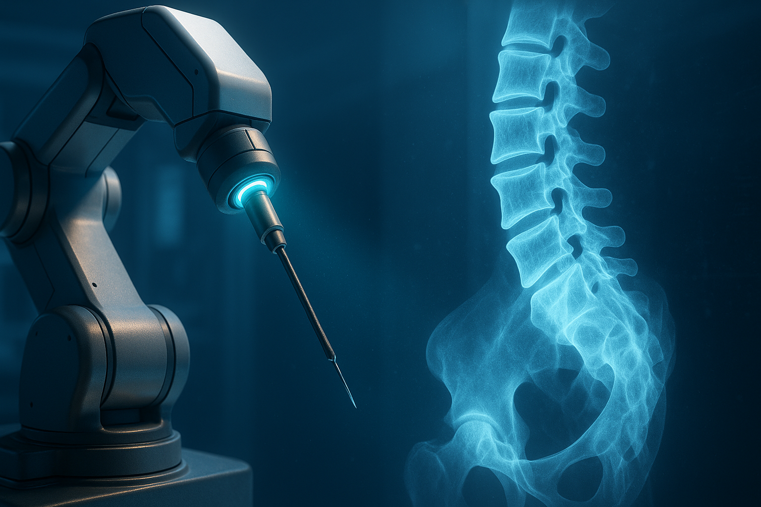
Surgical robotics has long promised precision beyond human hands. Yet, the real constraint has never been mechanics — it’s perception. In high-stakes fields like spinal surgery, machines can move with submillimeter accuracy, but they can’t yet see through bone. That’s what makes the Johns Hopkins team’s new study, Investigating Robot Control Policy Learning for Autonomous X-ray-guided Spine Procedures, quietly radical. It explores whether imitation learning — the same family of algorithms used in self-driving cars and dexterous robotic arms — can enable a robot to navigate the human spine using only X-ray vision.
The question sounds clinical. The implications are not. If robots can reason spatially from sparse, noisy, and indirect 2D images, they could extend automation into domains once thought unreachable without 3D sensors or CT scans. But as the study shows, this vision remains part aspiration, part cautionary tale.
Background — From imitation to autonomy
Imitation learning has made remarkable strides in the past few years. Architectures like Action Chunking with Transformers (ACT) and Surgical Robot Transformer (SRT) learn by example — predicting sequences of human-like actions directly from visual cues. In surgical contexts, these models can mimic a surgeon’s hand movements frame by frame, executing complex multi-step workflows.
But fluoroscopy-guided spine procedures present an entirely different challenge. Surgeons rely on bi-planar X-rays, which are 2D projections of an inherently 3D anatomy. Interpreting them requires both geometric reasoning and anatomical intuition — skills that take years of experience. Transferring that tacit expertise into a neural policy model is a formidable test of whether “seeing like a human” can ever be replaced by “learning like a machine.”
Analysis — Teaching robots to plan from shadows
The team developed a full in silico sandbox, simulating thousands of X-ray-guided cannula insertions with high anatomical realism. Using datasets derived from the New Mexico Decedent Image Database, they created a library of correct trajectories — effectively, the muscle memory of a skilled surgeon encoded into data.
A transformer-based imitation learning policy was then trained to adjust the tool’s position based only on dual-view (anterior-posterior and lateral) X-ray inputs. The model learned to predict incremental “delta actions” — fine-grained adjustments in translation, rotation, and depth — to guide the cannula safely into the pedicle.
| Metric | Synthetic Cases | Fractured Anatomy | Real X-rays |
|---|---|---|---|
| Acceptance Rate (Grades A+B) | 68.5% | 49.2% | 34.8% |
| Mean Entry Error | 5.46 mm | — | — |
| Mean Angular Offset | 3.53° | — | — |
The policy succeeded on the first attempt in over two-thirds of simulations — maintaining safe intra-pedicular trajectories across thoracic and lumbar vertebrae. Even on real bi-planar X-rays, trained purely in simulation, it managed plausible trajectories one-third of the time. That’s not surgical-grade reliability, but it’s an extraordinary proof of concept.
Findings — Where robots stumble
Performance dropped sharply in fractured anatomies, where geometry deviates from textbook cases. The system handled orientation well but often missed the entry point — a few millimeters off, but enough to risk cortical breach. It’s a revealing failure mode: the model can imitate precision, but without deeper anatomical priors, it doesn’t understand precision.
The ablation experiments confirmed this fragility. Remove one X-ray view, and success rates collapsed from 68% to below 20%. Even small perturbations in starting pose triggered cascading errors. The robot learned the “how” of insertion but not the “why.”
Implications — From CT-free surgery to human-AI hybrid navigation
If perfected, this kind of imitation-driven control could eliminate the need for intra-operative CT — reducing radiation, equipment cost, and setup time. It could also enable lightweight, portable robotic assistants that operate in low-resource hospitals.
Yet the ethical and safety implications are profound. X-rays provide sparse, high-stakes visual feedback. Each frame costs radiation exposure; each millimeter of error risks paralysis. This domain exposes the limits of current imitation learning — it performs adequately only where data density is high and physical consequences are low.
The next frontier will not be better transformers, but better priors — hybrid systems that integrate domain knowledge, probabilistic reasoning, and continual human oversight. Robots won’t replace the spine surgeon anytime soon. But they may soon become perceptive collaborators — ones that watch every move, learn continuously, and never blink under radiation.
Conclusion — Seeing through the noise
The Johns Hopkins study doesn’t hand us autonomous surgery. It hands us something subtler: a mirror reflecting where imitation learning stops being intelligence and starts being mimicry. Between the grainy shadows of an X-ray and the precision of a surgeon’s judgment lies the real challenge of AI — not learning to act, but learning to understand.
Cognaptus: Automate the Present, Incubate the Future.